What’s the diagnosis?
Scarring alopecia with tufted folliculitis

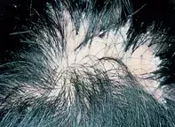
Figure 1. An irregular area of scarring alopecia on the vertex of the scalp.

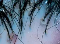
Figure 2. A closer view demonstrating tufted groups of hairs emerging from single follicular orifices and pustules.
Differential Diagnosis
Tinea capitis may present with pustules as well as broken hairs that appear as black dots on the scalp. Tufted follicles may occasionally be seen. Hair pluck and cultures will confirm the presence of tinea.
Dissecting cellulitis of the scalp occurs predominantly in young men with a background of nodulocystic acne. Fluctuant deep dermal and subcutaneous abscesses develop through the scalp, which may discharge via sinuses. The process is highly destructive and leads to extensive scarring.
Acne cheloidales is usually localised to the occipital area in men and is characterised by multiple nodulocystic lesions and follicular abscesses. The process is complicated by bacterial infection and hairs perforating the follicular canals because of poor grooming practices.
Tufted folliculitis (folliculitis decalvans) is the correct diagnosis. This is a pustular form of folliculitis due to Staphylococcus aureus. The disruption of the follicular canals results in their subsequent fusion to form common follicular openings with tufts of hair. Tufted folliculitis is seen most frequently as a component of a distinctive form of pustular scarring alopecia compounded, possibly, by delayed hypersensitivity reaction to S. aureus (folliculitis decalvans). This results in an expanding scar with peripheral pustules. Cultures and bacterial sensitivities are useful prior to treatment particularly in persistent cases. Treatment with long-term combinations of antistaphylococcal antibiotics such as cephalexin, erythromycin, rifampicin, sodium fusidate or ciprofloxacin may help to halt the process.
A 34-year-old woman gave an 18-month history of progressive hair loss over the vertex of her scalp (Figure 1). This was associated with tenderness and occasional pustules. Closer examination showed scarring of the scalp with loss of follicular orifices. There were also multiple tufts of dark hair emerging from single openings (Figure 2).

