What’s the diagnosis?
Itchy coin-shaped lesions

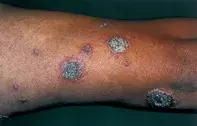
Figure 1. Crusted coin-shaped lesions with small peripheral vesicles on the patient’s limb.

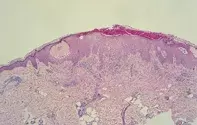
Figure 2. Skin biopsy showing locules of pink serum in the epidermis and prominent inflammation in the upper dermis.
Differential Diagnosis
Psoriasis is usually not pruritic and usually does not have a crusted appearance or vesicles. Skin biopsy in psoriasis shows a thick epidermis with neutrophil microabscesses.
Impetigo is due to superficial cutaneous infection with staphylococci or streptococci. It has an identical crusted appearance to that seen in this case, but it lacks the rather symmetrical pattern and the pruritic vesicles.
Tinea may present with crusted lesions, but often these are associated with central clearing and peripheral pustules. Skin biopsy and cultures should reveal fungal elements.
Secondary syphilis may rarely present as markedly crusted lesions, but these are usually not pruritic. The presence of generalised symptoms, lymphadenopathy and positive serological test results should help to confirm the diagnosis.
Nummular (discoid) dermatitis is the correct diagnosis and has a predilection for the limbs but may extend to the trunk. This dermatitis may complicate stasis eczema. Bacterial infection is an important element and may trigger extension of other lesions. Allergy patch testing may be helpful in chronic cases. Nummular dermatitis usually will respond only when a combination of topical corticosteroids and systemic antibiotics, to deal with staphylococci, are employed. Moisturisers may be required for associated dry skin.
Over a three-month period, a 43-year-old man developed numerous round, crusted lesions on his limbs that were extremely pruritic (Figure 1). The individual lesions varied in size from a few millimetres to 2 to 3 cm in diameter and had a vesicular border. There was no history of atopy. Skin biopsy showed an epidermis that was associated with locules of serum and a marked inflammatory reaction that extended into the upper dermis (Figure 2).

