What’s the diagnosis?
Intermittently itchy reddish-brown spots

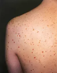
Figure 1. Reddish-brown lesions on the patient’s back and upper limbs.

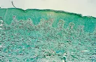
Figure 2. A skin biopsy demonstrating numerous cells in the dermis that stain as mast cells (chloroacetate esterase stain).
Differential Diagnosis
Multiple lentigines or junctional naevi may have a similar appearance but usually are more deeply pigmented and do not progressively increase in number over a short period in this age group. Skin biopsy of naevi shows junctional and dermal nests of melanocytes as well as melanin pigment.
Vasculitis may be associated with progressive purpuric lesions that are admixed with brown haemosiderin pigment. The individual lesions resolve and are usually concentrated on dependent areas rather than more diffusely distributed. Skin biopsy shows vascular necrosis with leucocytoclasis.
Eruptive histiocytomas represent a rare condition associated with red to flesh-coloured papules that may be widespread and last for several years. Skin biopsy shows a dermal infiltrate composed of sheets of histiocytes that usually lack a granular cytoplasm.
Mastocytosis (urticaria pigmentosa) is the correct diagnosis and may develop at all ages, particularly in childhood. Systemic mastocytosis, involving the bone marrow, liver, spleen, gastrointestinal tract or lymph nodes, may develop in up to 40% of adults. Release of mast cell mediators, including histamine, leukotrienes and other prostaglandin derivatives, may produce itching, wheezing, flushing and tachycardia as well as hypotensive episodes. Nausea, abdominal pain and diarrhoea may also occur. Avoidance of mast cell activators, such as heat, aspirin and related compounds as well as some forms of radiographic dyes, is important as these may result in anaphylaxis. At present there is no effective means of treating mast cell disease and the process usually remains indolent but may transform to a mast cell leukaemia or be associated with myeloid leukaemia.
Over a three-year period, a 32-year-old man noted an increasing number of 1 to 3 mm reddish-brown spots on his trunk and limbs (Figure 1). The face, palms and soles were relatively spared. The individual lesions had remained stable but persistent. Occasionally an urticarial reaction developed around a lesion that had been rubbed. A skin biopsy revealed an extensive infiltrate of mononuclear cells with fine granular cytoplasm. The cells showed positivity with chloroacetate esterase stain (Figure 2).

